ARRS Case of the Week
MUSCULOSKELETAL IMAGING
Case Authors: Daniel E. Wessell, MD, PhD, Mayo Clinic Florida
History
20-year-old man with midfoot pain and swelling after trauma; findings on initial non–weight-bearing radiographs in the emergency department are normal; the next imaging step must be determined.
Findings
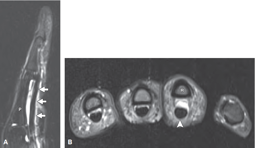
Non–weight-bearing dorsoplantar radiograph of the foot (A) shows normal alignment between the medial cuneiform and the second metatarsal base (arrow). No fracture is visible. Subsequently obtained weight-bearing dorsoplantar radiograph of the foot (B) shows widening of the joint space (arrowhead) between the medial cuneiform and the second metatarsal base. Long‑axis T2-weighted fat-suppressed MR image of the midfoot (C) shows disruption of the ligament (arrow) between the medial cuneiform (M) and the second metatarsal base (2) with edema in the adjacent soft tissues.
This page is updated with new content weekly. It was last updated on August 8, 2022.
You May Also Be Interested In